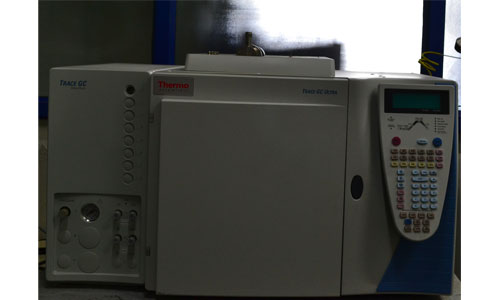
Facility is integrated with a methanator for detection and evaluation of trace amounts of CO and CO2 gases present in the reactant/ product stream of gases
Gas chromatograph with TCD, FID and FPD detectors is used to analyse the exhaust gas from the fuel cell especially, when the fuel cell is operated with methanol and reformate gas.
Centre for Fuel Cell Technology (CFCT)
Dr. N. Rajalakshmi
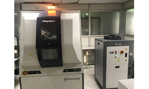
The high-resolution X-ray diffractometer provided with SmartLab Guidance software, acts as an intelligent interface for the user. The system incorporates a high resolution θ/θ closed loop goniometer drive system, cross beam optics (CBO), an in-plane scattering arm, and an optional 3.0 kW rotating anode generator. High flexibility by coupling a computer controlled alignment system with a fully automated optical system makes it easy to switch between hardware modes. Optional in-plane diffraction arm for in-plane measurements without reconfiguration and focusing and parallel beam geometries without reconfiguration.
Centre for Fuel Cell Technology (CFCT)
Dr. N. Rajalakshmi
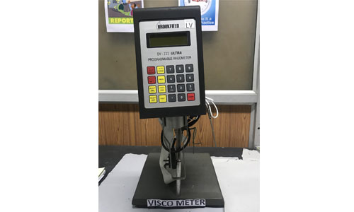
This Rheometer easily predict a material's flow, spray, or pumping behavior by studying shear rate profiles and has continuous display of: Viscosity (cP or mPa·s), Temperature (°C or °F), Shear Rate, Shear Stress, % Torque, Spindle, Program status 2600 speeds for incredible characterization possibilities It has easy-to-use keypad with numeric keys for stand-alone program entry.
Centre for Fuel Cell Technology (CFCT)
Dr. N. Rajalakshmi
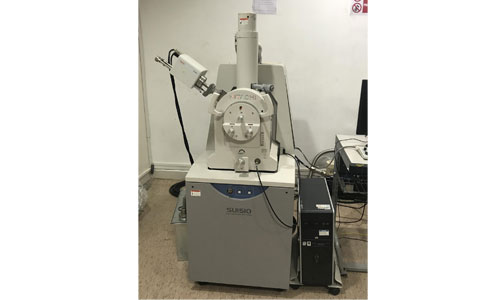
Medium size chamber variable pressure SEM with Quad Bias gun electronics which improves low voltage performance and increases beam the current. Dual high-take-off ports accommodate two EDS detectors mounted 180 degrees apart to tice the analytical data collection plus eliminate X-ray map shadows associated with rough sample surfaces. A high speed, clean, efficient TMP eliminates the need for water cooling. Advantages of being compact, high performance and user friendly. For quick observation of non-conductive samples the SU1510 utilizes variable pressure mode that eliminates negative charging, and provides the optimum conditions for both imaging and Energy Dispersive X-ray microanalysis. The specimen chamber and stage have been designed to accommodate samples as large as 153 mm in diameter. Simultaneous EDX microanalysis and imaging can be completed on a sample that is up to 60mm in height at the analytical working distance of 15 mm.
Centre for Materials Characterization and Testing (CMCT)
Dr. N. Rajalakshmi
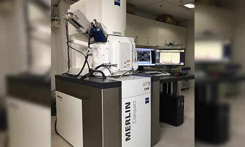
High resolution Field Emission Gun scanning electron microscope (FEG-SEM), (Model: Merlin Compact, Make: Zeiss, Germany) is available at ARCI, Chennai. The instrument has a best resolution of 0.8 nm with accelerating voltage variable from 0.02 to 30 keV. The FEG source is of thermionic emission type. The SEM is equipped with Secondary electron detector, In-lens detector, Back scattered Electron detector and EDS detector (EDAX, USA) for compositional analysis. The sample stage is a 5 axis euccentric stage with a tilt angle ranging from -3 to 70 0. The Gemini I column enables high resolution and high probe current making it more suitable for analytical applications.
Centre for Automotive Energy Materials (CAEM)
Dr. D Prabhu
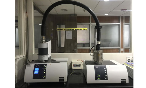
It consists of a vacuum-tight thermo micro balance system, microprocessor-controlled, with integrated calibration weight, with water-cooled heater, exchangeable sample carrier including radiation shield, programmable gas flow for 3 gases and integrated heating power supply, automated lid lifting device (prepared autovac for automated vacuum generation/ventilation). This instrument comes with QMS coupling for evolved gas analysis from 1-300 amu and a processing module of the Aeolos software (as of version 7.02), enabling a triggered start and stop as well as the transfer of the temperature signal into the Aeolos software for programming and control. unit for data acquisition and processor of max. 64 measuring channels (mass and mass range resp.)
Centre for Fuel Cell Technology (CFCT)
Dr. N. Rajalakshmi
